Tuesday, September 13, 2011

ALL ABOUT AMINO ACID METABOLISM

DIGESTION OF DIETARY PROTIENS
Digestion of dietary proteins has two principle sites, which are gastric digestion and deudenal digestion but it usually starts in the stomach. Secretion of HCl activates formation of pepsin from pepsinogen which hydrolyses peptide bonds of dietary proteins to release aromatic amino acids. Similarly, in the small intestine digestion continues. Zymogens are activated for the breakdown of peptide bonds to release amino acids which are absorbed into the small intestine.
AMINO ACID DEAMINATION
Transamination is a process by which amino group is transferred to the keto acid to yield the keto acid of the original amino acid and a new amino acid, catalyzed by amino transferases. Glutamate is oxidatively deaminated in the mitochondrion and release ammonia.
THE UREA CYCLE
This cyclic pathway takes place in the liver, partly in the mitochondria and partly in cytosol. There is interrelation between urea cycle and citric acid cycle. Fumarate of the urea cycle is taken up by citric acid cycle. Carbamoyl phosphate synthetase I is the regulatory enzyme.
METABOLIC BREAK DOWN OF INDIVIDUAL AMINO ACID
Catabolism of amino acids gives rise to the intermediate compounds of citric acid cycle. Alanine, serine, cysteine and asparagine are converted to oxaloacetate. Glutamine, proline, arginine and histidine are converted to α- ketoglutarate through glutamate. Succinyl CoA is a point of entry for non polar amino acids like methionine, valine and isoleucine. Leucine is degraded to acetyl CoA and acetoacetate. Tryptophan, lysine, leucine, phenylalanine, tyrosine and isoleucine donate their carbons to acetyl CoA. Various disorders of amino acid catabolism are observed due to the defective enzymes. Tryptophan, tyrosine, glycine and glutamate are the precursors of some of the biologically important compounds. Biotin, Tetrahydrofolate or S-Adenosyl methionine is the enzyme cofactors in catabolism which transfer carbon compounds.
AMINO ACID BIOSYNTHESIS
Nonessential amino acids are formed from intermediates of carbohydrate metabolism. Alanine is formed from pyruvate and aspartate from oxaloacetate. Asparagine is formed from aspartate. Glutamate is formed from α ketoglutarate and glutamine from glutamate. Glutamate is the precursor of proline and arginine. Cysteine is synthesized from 3 – phosphoglycerate. During the process, serine is the intermediate compound which gives rise to glycine. Threonine is an essential amino acid. It is formed from β-aspartate. Methionine and lysine alsohave a common precursor. Valine, Leucine and isoleucine are formed from pyruvate. Phenyl alanine, tyrosine and Tryptophan are formed from phosphoenol pyruvate and erythrose – 4 – phosphate through the intermediate compound called chorismate. Histidine usually form L-glutamate.

Saturday, September 10, 2011

All you need to know about ovulation

OVULATION Ovulation is the shadding of the ovum from the ovary, Also is the process in a female's menstrual cycle by which a mature ovarian follicle ruptures and discharges an ovum (also known as an oocyte, female gamete, or casually, an egg). Ovulation also occurs in the estrous cycle of other female mammals, which differs in many fundamental ways from the menstrual cycle. The time immediately surrounding ovulation is referred to as the ovulatory phase or the periovulatory period. overview The process of ovulation is controlled by the hypothalamus of the brain and through the release of hormones secreted in the anterior lobe of the pituitary gland, luteinizing hormone (LH) and follicle- stimulating hormone (FSH). In the pre-ovulatory phase of the menstrual cycle, the ovarian follicle will undergo a series of transformations called cumulus expansion, which is stimulated by FSH. After this is done, a hole called the stigma will form in the follicle, and the ovum will leave the follicle through this hole. Ovulation is triggered by a spike in the amount of FSH and LH released from the pituitary gland. During the luteal (post-ovulatory) phase, the ovum will travel through the fallopian tubes toward the uterus. If fertilized by a sperm, it may perform implantation there 6–12 days later. Ovulation occurs when a mature egg is released from the ovary into the abdominal cavity.[1] Afterwards, it will eventually become available to be fertilized. Concomitantly, the lining of the uterus is thickened to be able to receive a fertilized egg. If no conception occurs, the uterine lining as well as blood will be shed in menstruation. In humans, the few days near ovulation constitute the fertile phase. The time from the beginning of the last menstrual period (LMP) until ovulation is, on average, 14.6[2] days, but with substantial variation both between women and between cycles in any single woman, with an overall 95% prediction interval of 8.2 to 20.5[2] days. Cycle length alone is not a reliable indicator of the day of ovulation. While in general an earlier ovulation will result in a shorter menstrual cycle, and vice versa, the luteal (post-ovulatory) phase of the menstrual cycle may vary by up to a week between women.o as the ovulatory phase or the periovulatory period. overview The process of ovulation is controlled by the hypothalamus of the brain and through the release of hormones secreted in the anterior lobe of the pituitary gland, luteinizing hormone (LH) and follicle- stimulating hormone (FSH). In the pre-ovulatory phase of the menstrual cycle, the ovarian follicle will undergo a series of transformations called cumulus expansion, which is stimulated by FSH. After this is done, a hole called the stigma will form in the follicle, and the ovum will leave the follicle through this hole. Ovulation is triggered by a spike in the amount of FSH and LH released from the pituitary gland. During the luteal (post-ovulatory) phase, the ovum will travel through the fallopian tubes toward the uterus. If fertilized by a sperm, it may perform implantation there 6–12 days later. Ovulation occurs when a mature egg is released from the ovary into the abdominal cavity.[1] Afterwards, it will eventually become available to be fertilized. Concomitantly, the lining of the uterus is thickened to be able to receive a fertilized egg. If no conception occurs, the uterine lining as well as blood will be shed in menstruation. In humans, the few days near ovulation constitute the fertile phase. The time from the beginning of the last menstrual period (LMP) until ovulation is, on average, 14.6[2] days, but with substantial variation both between women and between cycles in any single woman, with an overall 95% prediction interval of 8.2 to 20.5[2] days. Cycle length alone is not a reliable indicator of the day of ovulation. While in general an earlier ovulation will result in a shorter menstrual cycle, and vice versa, the luteal (post-ovulatory) phase of the menstrual cycle may vary by up to a week between women.

Wednesday, September 7, 2011

MECHANISM OF PANCREATIC SECRETION.

The pancreatic enzymes are synthesized in ribosomes, which are attached to the endoplasmic recticulum of acinar cells in pancrease. The raw materials for synthesis of pancreatic enzymes are the amino acids which are derived from blood. After synthesis, the enzymes are packed into different zymogen granules by golgi apparatus and stored in cytoplasm. When stimulated, the acinar cells release the zymogen granules into the pancreatic duct. From the granules, the enzymes are liberated into intestine. ic enzymes are synthesized in ribosomes, which are attached to the endoplasmic recticulum of acinar cells in pancrease. The raw materials for synthesis of pancreatic enzymes are the amino acids which are derived from blood. After synthesis, the enzymes are packed into different zymogen granules by golgi apparatus and stored in cytoplasm. When stimulated, the acinar cells release the zymogen granules into the pancreatic duct. From the granules, the enzymes are liberated into intestine .

Wednesday, May 18, 2011

RENAL FUNCTION TESTS II

Kidney function tests

Kidney function tests are common lab tests used to evaluate how well the kidneys are working. See links below for details of how each test is performed:

1.BUN

BUN stands for blood urea nitrogen. Urea nitrogen is what forms when protein breaks down.
A test can be done to measure the amount of urea nitrogen in the blood.

How the Test is Performed

Blood is typically drawn from a vein, usually from the inside of the elbow or the back of the hand. The site is cleaned with germ-killing medicine (antiseptic). The health care provider wraps an elastic band around the upper arm to apply pressure to the area and make the vein swell with blood.
Next, the health care provider gently inserts a needle into the vein. The blood collects into an airtight vial or tube attached to the needle. The elastic band is removed from your arm.
Once the blood has been collected, the needle is removed, and the puncture site is covered to stop any bleeding.
In infants or young children, a sharp tool called a lancet may be used to puncture the skin and make it bleed. The blood collects into a small glass tube called a pipette, or onto a slide or test strip. A bandage may be placed over the area if there is any bleeding.

How to Prepare for the Test

Many drugs affect BUN levels. Before having this test, make sure the health care provider knows which medications you are taking.
Drugs that can increase BUN measurements include:
  • Allopurinol
  • Aminoglycosides
  • Amphotericin B
  • Aspirin (high doses)
  • Bacitracin
  • Carbamazepine
  • Cephalosporins
  • Chloral hydrate
  • Cisplatin
  • Colistin
  • Furosemide
  • Gentamicin
  • Guanethidine
  • Indomethacin
  • Methicillin
  • Methotrexate
  • Methyldopa
  • Neomycin
  • Penicillamine
  • Polymyxin B
  • Probenecid
  • Propranolol
  • Rifampin
  • Spironolactone
  • Tetracyclines
  • Thiazide diuretics
  • Triamterene
  • Vancomycin
Drugs that can decrease BUN measurements include:
  • Chloramphenicol
  • Streptomycin

How the Test Will Feel

When the needle is inserted to draw blood, some people feel moderate pain, while others feel only a prick or stinging sensation. Afterward, there may be some throbbing.

Why the Test is Performed

The BUN test is often done to check kidney function.

Normal Results

7 - 20 mg/dL. Note that normal values may vary among different laboratories.

What Abnormal Results Mean

Higher-than-normal levels may be due to:
  • Heart attack
  • Kidney disease,
Lower-than-normal levels may be due to:
  • Liver failure
  • Low protein diet
  • Malnutrition
  • Over-hydration
Additional conditions under which the test may be done include:
  • Acute nephritic syndrome
  • Atheroembolic kidney disease
  • Diabetic nephropathy/sclerosis
  • Epilepsy
  • Goodpasture syndrome
  • Hepatokidney syndrome
  • Lupus nephritis
  • Malignant hypertension (arteriolar nephrosclerosis)
  • Secondary systemic amyloidosis
  • Wilms' tumor

Risks

Veins and arteries vary in size from one patient to another and from one side of the body to the other. Obtaining a blood sample from some people may be more difficult than from others.
Other risks are slight but may include:
  • Excessive bleeding
  • Fainting or feeling light-headed
  • Hematoma (blood accumulating under the skin)
  • Infection (a slight risk any time the skin is broken)

Considerations

For people with liver diseases the BUN level may be low even if the kidneys are normal.

Alternative Names

Blood urea nitrogen

2.Creatinine - blood

Creatinine is a breakdown product of creatine, which is an important part of muscle. This article discusses the laboratory test to measure the amount of creatinine in the blood.

How the Test is Performed

Blood is drawn from a vein, usually from the inside of the elbow or the back of the hand. The site is cleaned with germ-killing medicine (antiseptic). The health care provider wraps an elastic band around the upper arm to apply pressure to the area and make the vein swell with blood.
Next, the health care provider gently inserts a needle into the vein. The blood collects into an airtight vial or tube attached to the needle. The elastic band is removed from your arm.
Once the blood has been collected, the needle is removed, and the puncture site is covered to stop any bleeding.
In infants or young children, a sharp tool called a lancet may be used to puncture the skin and make it bleed. The blood collects into a small glass tube called a pipette, or onto a slide or test strip. A bandage may be placed over the area if there is any bleeding.

How to Prepare for the Test

The health care provider may tell you to stop taking certain drugs that may affect the test. Such drugs include:
  • Aminoglycosides (for example, gentamicin)
  • Cimetidine
  • Heavy metal chemotherapy drugs (for example, Cisplatin)
  • Kidney damaging drugs such as cephalosporins (for example, cefoxitin)
  • Trimethoprim

How the Test Will Feel

When the needle is inserted to draw blood, some people feel moderate pain, while others feel only a prick or stinging sensation. Afterward, there may be some throbbing.

Why the Test is Performed

The test is done to evaluate kidney function. Creatinine is removed from the body entirely by the kidneys. If kidney function is abnormal, creatinine levels will increase in the blood (because less creatinine is released through your urine).
Creatinine levels also vary according to a person's size and muscle mass.

Normal Results

A normal value is 0.8 to 1.4 mg/dL.
Females usually have a lower creatinine than males, because they usually have less muscle mass.
Note: Normal value ranges may vary slightly among different laboratories. Talk to your doctor about the meaning of your specific test results.

The Kidneys and How They Works

.
What do the kidneys do?
The kidneys are bean-shaped organs, each about the size of a fist. They are located near the middle of the back, just below the rib cage, one on each side of the spine. The kidneys are sophisticated reprocessing machines. Every day, a person’s kidneys process about 200 quarts of blood to sift out about 2 quarts of waste products and extra water. The wastes and extra water become urine, which flows to the bladder through tubes called ureters. The bladder stores urine until releasing it through urination.
Drawing of the urinary tract in a male figure with labels for the kidneys, bladder, and ureters.
The kidneys remove wastes and water from the blood to form urine. Urine flows from the kidneys to the bladder through the ureters.
Wastes in the blood come from the normal breakdown of active tissues, such as muscles, and from food. The body uses food for energy and self-repairs. After the body has taken what it needs from food, wastes are sent to the blood. If the kidneys did not remove them, these wastes would build up in the blood and damage the body.
The actual removal of wastes occurs in tiny units inside the kidneys called nephrons. Each kidney has about a million nephrons. In the nephron, a glomerulus—which is a tiny blood vessel, or capillary—intertwines with a tiny urine-collecting tube called a tubule. The glomerulus acts as a filtering unit, or sieve, and keeps normal proteins and cells in the bloodstream, allowing extra fluid and wastes to pass through. A complicated chemical exchange takes place, as waste materials and water leave the blood and enter the urinary system.
Drawing of a kidney. Labels show where blood with wastes enter the kidney, clean blood leaves the kidney, and wastes (urine) are sent to the bladder. An inset shows a microscopic view of a nephron. Labels point to the glomerulus and the tubule.
In the nephron (left), tiny blood vessels intertwine with urine-collecting tubes. Each kidney contains about 1 million nephrons.
At first, the tubules receive a combination of waste materials and chemicals the body can still use. The kidneys measure out chemicals like sodium, phosphorus, and potassium and release them back to the blood to return to the body. In this way, the kidneys regulate the body’s level of these substances. The right balance is necessary for life.
In addition to removing wastes, the kidneys release three important hormones:
  • erythropoietin, or EPO, which stimulates the bone marrow to make red blood cells
  • renin, which regulates blood pressure
  • calcitriol, the active form of vitamin D, which helps maintain calcium for bones and for normal chemical balance.

RENAL FUNCTION TEST I


Creatinine Clearance CCr

The creatinine clearance is not widely done any more, due to the difficulty in assuring a complete urine collection. When doing such a determination, to assess the adequacy of a complete collection, one always calculates the amount of creatinine excreted over a 24-hour period. This amount varies with muscle mass, and is higher in young people vs. old, in blacks vs. whites, and in men vs. women. An unexpectedly low or high 24-hour creatinine excretion rate voids the test. Nevertheless, in cases where estimates of creatinine clearance from serum creatinine are unreliable, creatinine clearance remains a useful test. These cases include "estimation of GFR in individuals with variation in dietary intake (vegetarian diet, creatine supplements) or muscle mass (amputation, malnutrition, muscle wasting), since these factors are not specifically taken into account in prediction equations.

 Estimated values

eC_{Cr} = \frac { \mbox{(140 - Age)} \ \times \ \mbox{Mass (in kilograms)} \ \times \ [{0.85\ if\ Female}]} {\mbox{72} \ \times \ \mbox{Serum Creatinine (in mg/dL)}}
This formula expects weight to be measured in kilograms and creatinine to be measured in mg/dL, as is standard in the USA. The resulting value is multiplied by a constant of 0.85 if the patient is female. This formula is useful because the calculations are simple and can often be performed without the aid of a calculator
When serum creatinine is measured in µmol/L:
eC_{Cr} = \frac { \mbox{(140 - Age)} \ \times \ \mbox{Mass (in kilograms)} \ \times \ {Constant} } {\mbox{Serum Creatinine (in } \mu \mbox{mol/L)}}
Where Constant is 1.23 for men and 1.04 for women.
One interesting feature of the Cockcroft and Gault equation is that it shows how dependent the estimation of CCr is based on age. The age term is (140 - age). This means that a 20-year-old person (140-20 = 120) will have twice the creatinine clearance as an 80-year-old (140-80 = 60) for the same level of serum creatinine (120 is twice as great as 60). The C-G equation also shows that a woman will have a 15% lower creatinine clearance than a man at the same level of serum creatinine.

 Estimated GFR (eGFR) using Modification of Diet in Renal Disease (MDRD)

\mbox{eGFR} = \mbox{186}\ \times \ \mbox{Serum Creatinine}^{-1.154} \ \times \ \mbox{Age}^{-0.203} \ \times \ {[1.212\ if\ Black]} \ \times \ {[0.742\ if\ Female]}
For creatinine in µmol/L:
\mbox{eGFR} = \mbox{32788}\ \times \ \mbox{Serum Creatinine}^{-1.154} \ \times \ \mbox{Age}^{-0.203} \ \times \ {[1.212\ if\ Black]} \ \times \ {[0.742\ if\ Female]}
Creatinine levels in µmol/L can be converted to mg/dL by dividing them by 88.4. The 32788 number above is equal to 186×88.41.154.
\mbox{eGFR} = \mbox{170}\ \times \ \mbox{Serum Creatinine}^{-0.999} \ \times \ \mbox{Age}^{-0.176} \ \times \ {[0.762\ if\ Female]} \ \times \ {[1.180\ if\ Black]} \ \times \ \mbox{BUN}^{-0.170} \ \times \ \mbox{Albumin}^{+0.318}
Where the creatinine and blood urea nitrogen concentrations are both in mg/dL. The albumin concentration is in g/dL.
These MDRD equations are to be used only if the laboratory has NOT calibrated its serum creatinine measurements to isotope dilution mass spectroscopy (IDMS). When IDMS-calibrated serum creatinine is used (which is about 6% lower), the above equations should be multiplied by 175/186 or by 0.94086.
Since these formulae do not adjust for body mass, they (relative to the Cockcroft-Gault formula) underestimate eGFR for heavy people and overestimate it for underweight people. (see Cockcroft-Gault formula above)
Estimated GFR (eGFR) using the CKD-EPI formula
The CKD-EPI (Chronic Kidney Disease Epidemiology Collaboration) formula was published in May 2009. It was developed in an effort to create a formula more accurate than the MDRD formula, especially when actual GFR is greater than 60 mL/min per 1.73 m2.
Researchers pooled data from multiple studies to develop and validate this new equation. They used 10 studies that included 8254 participants, randomly using 2/3 of the data sets for development and the other 1/3 for internal validation. Sixteen additional studies, which included 3896 participants, were used for external validation.
The CKD-EPI equation performed better than the MDRD (Modification of Diet in Renal Disease Study) equation, especially at higher GFR, with less bias and greater accuracy. When looking at NHANES (National Health and Nutrition Examination Survey) data, the median estimated GFR was 94.5 mL/min per 1.73 m2 vs. 85.0 mL/min per 1.73 m2, and the prevalence of chronic kidney disease was 11.5% versus 13.1%.
The CKD-EPI equation, expressed as a single equation, is:
\mbox{eGFR} = \mbox{141}\ \times \ \mbox{min(SCr/k,1)}^{a} \ \times \ \mbox{max(SCr/k,1)}^{-1.209} \ \times \ \mbox{0.993}^{Age} \ \times \ {[1.018\ if\ Female]} \ \times \ {[1.159\ if\ Black]} \
where SCr is serum creatinine (mg/dL), k is 0.7 for females and 0.9 for males, a is -0.329 for females and -0.411 for males, min indicates the minimum of SCr/k or 1, and max indicates the maximum of SCr/k or 1.
A clearer version may be as follows: For creatinine (IDMS calibrated) in mg/dL:
African American Female
If serum creatinine (Scr) <= 0.7
\mbox{eGFR} = \mbox{166}\ \times \ \mbox{(SCr/0.7)}^{-0.329} \ \times \ \mbox{0.993}^{Age} \
If serum creatinine (Scr) > 0.7
\mbox{eGFR} = \mbox{166}\ \times \ \mbox{(SCr/0.7)}^{-1.209} \ \times \ \mbox{0.993}^{Age} \
African American Male
If serum creatinine (Scr) <= 0.9
\mbox{eGFR} = \mbox{163}\ \times \ \mbox{(SCr/0.9)}^{-0.411} \ \times \ \mbox{0.993}^{Age} \
If serum creatinine (Scr) > 0.9
\mbox{eGFR} = \mbox{163}\ \times \ \mbox{(SCr/0.9)}^{-1.209} \ \times \ \mbox{0.993}^{Age} \
White or other race Female
If serum creatinine (Scr) <= 0.7
\mbox{eGFR} = \mbox{144}\ \times \ \mbox{(SCr/0.7)}^{-0.329} \ \times \ \mbox{0.993}^{Age} \
If serum creatinine (Scr) > 0.7
\mbox{eGFR} = \mbox{144}\ \times \ \mbox{(SCr/0.7)}^{-1.209} \ \times \ \mbox{0.993}^{Age} \
White or other race Male
If serum creatinine (Scr) <= 0.9
\mbox{eGFR} = \mbox{141}\ \times \ \mbox{(SCr/0.9)}^{-0.411} \ \times \ \mbox{0.993}^{Age} \
If serum creatinine (Scr) > 0.9
\mbox{eGFR} = \mbox{141}\ \times \ \mbox{(SCr/0.9)}^{-1.209} \ \times \ \mbox{0.993}^{Age} \
\mbox{eGFR} = \mbox{exp}{(1.911+ 5.249/{Serum\ Creatinine} - 2.114/{Serum\ Creatinine}^2 - 0.00686 \ \times \ \mbox{Age} - {[0.205\ if\ Female]})}
If Serum Creatinine < 0.8 mg/dL, use 0.8 mg/dL for Serum Creatinine

Estimated GFR for children using Schwartz formula

In children, the Schwartz formula is used.This employs the serum (mg/dL), the child's height (cm) and a constant to estimate the glomerular filtration rate:
\mbox{eGFR} = \frac{ {k} \times {Height} }{Serum\ Creatinine}
Where k is a constant that depends on muscle mass, which itself varies with a child's age:
In first year of life, for pre-term babies K=0.33 and for full-term infants K=0.45
For infants and children of age 1 to 12 years, K=0.55
The method of selection of the K-constant value has been questioned as being dependent upon the gold-standard of renal function used (i.e., creatinine clearance, inulin clearance, etc.) and also may be dependent upon the urinary flow rate at the time of measurement
In 2009, the formula was updated to use standardized serum creatinine (recommend k=0.413) and additional formulas that allow improved precision were derived if serum cystatin measured in addition to serum creatinine importance of calibration of the serum creatinine level and the IDMS standardization effort

One problem with any creatinine-based equation for GFR is that the methods used to assay creatinine in the blood differ widely in their susceptibility to non-specific chromogens, which cause the creatinine value to be overestimated. In particular, the MDRD equation was derived using serum creatinine measurements that had this problem. The NKDEP program in the United States has attempted to solve this problem by trying to get all laboratories to calibrate their measures of creatinine to a "gold standard", which in this case is isotope dilution mass spectroscopy (IDMS). At the present time in late 2009 not all labs in the U.S. have changed over to the new system. There are two forms of the MDRD equation that are available, depending on whether or not creatinine was measured by an IDMS-calibrated assay. The CKD-EPI equation is designed to be used with IDMS-calibrated serum creatinine values only.

Cystatin C

Problems with creatinine (varying muscle mass, recent meat ingestion, etc.) have led to evaluation of alternative agents for estimation of GFR. One of these is, a ubiquitous protein secreted by most cells in the body (it is an inhibitor of cysteine protease).
Cystatin C is freely filtered at the glomerulus. After filtration, Cystatin C is reabsorbed and catabolized by the tubular epithelial cells, with only small amounts excreted in the urine. Cystatin C levels are therefore measured not in the urine, but in the bloodstream.
Equations have been developed linking estimated GFR to serum cystatin C levels. Most recently, some proposed equations have combined creatinine and cystatine
Normal ranges
The normal range of GFR, adjusted for is similar in men and women, and is in the range of 100-130 ml/min/1.73m2. In children, GFR measured by inulin clearance remains close to about 110 ml/min/1.73m2 down to about 2 years of age in both sexes, and then it progressively decreases. After age 40, GFR decreases progressively with age, by about 0.4 - 1.2 mL/min per year.

Chronic kidney disease stages 

Risk factors for kidney disease include diabetes, high blood pressure, family history, older age, ethnic group and smoking. For most patients, a GFR over 60 mL/min/1.73m2 is adequate. But significant decline of the GFR from a previous test result can be an early indicator of kidney disease requiring medical intervention. The sooner kidney dysfunction is diagnosed and treated the greater odds of preserving remaining nephrons, and preventing the need for dialysis.

The (CKD) is described by six stages; the most severe three are defined by the MDRD-eGFR value, and first three also depend on whether there is other evidence of kidney disease
0) Normal kidney function – GFR above 90mL/min/1.73m2
1) CKD1 – GFR above 90mL/min/1.73m2 with evidence of kidney damage
2) CKD2 (Mild) – GFR of 60 to 89 mL/min/1.73m2 with evidence of kidney damage
3) CKD3 (Moderate) – GFR of 30 to 59 mL/min/1.73m2
4) CKD4 (Severe) – GFR of 15 to 29 mL/min/1.73m2
5) CKD5 Kidney failure - GFR less than 15 mL/min/1.73m2 Some people add CKD5D for those stage 5 patients requiring dialysis; many patients in CKD5 are not yet on dialysis.
Note: others add a "T" to patients who have had a transplant regardless of stage.
Not all clinicians agree with the above classificaiton, suggesting that it may overlabel patients with mildly reduced kidney function, especially the elderly, as having a disease. A conference was held in 2009 regarding these controversies by Kidney Disease: Improving Global Outcomes (KDIGO) on CKD: Definition, Classification and Prognosis, gathering data on CKD prognosis to refine the definition and staging of CKD.

Saturday, April 30, 2011

Control of RespiratioN

Your respiratory rate changes. When active, for example, your respiratory rate goes up; when less active, or sleeping, the rate goes down. Also, even though the respiratory muscles are voluntary, you can't consciously control them when you're sleeping. So, how is respiratory rate altered & how is respiration controlled,
  • controls automatic breathing
  • consists of interacting neurons that fire either during inspiration (I neurons) or expiration (E neurons)
    • I neurons - stimulate neurons that innervate respiratory muscles (to bring about inspiration)
    • E neurons - inhibit I neurons (to 'shut down' the I neurons & bring about expiration)
Apneustic center (located in the pons) - stimulate I neurons (to promote inspiration) Pneumotaxic center (also located in the pons) - inhibits apneustic center & inhibits inspiration
 

Factors involved in increasing respiratory rate
  • Chemoreceptors - located in aorta & carotid arteries (peripheral chemoreceptors) & in the medulla (central chemoreceptors)
  • Chemoreceptors (stimulated more by increased CO2 levels than by decreased O2 levels) > stimulate Rhythmicity Area > Result = increased rate of respiration


Heavy exercise ==> greatly increases respiratory rate
Mechanism?
  • NOT increased CO2
  • Possible factors:
    • reflexes originating from body movements (proprioceptors)
    • increase in body temperature
    • epinephrine release (during exercise)
  • impulses from the cerebral cortex (may simultaneously stimulate rhythmicity area & motor neurons).

Hemoglobin saturation RATE

  • extent to which the hemoglobin in blood is combined with O2
  • depends on PO2 of the blood:


The relationship between oxygen levels and hemoglobin saturation is indicated by the oxygen-hemoglobin dissociation (saturation) curve (in the graph above). You can see that at high partial pressures of O2 (above about 40 mm Hg), hemoglobin saturation remains rather high (typically about 75 - 80%). This rather flat section of the oxygen-hemoglobin dissociation curve is called the 'plateau.'
Recall that 40 mm Hg is the typical partial pressure of oxygen in the cells of the body. Examination of the oxygen-hemoglobin dissociation curve reveals that, under resting conditions, only about 20 - 25% of hemoglobin molecules give up oxygen in the systemic capillaries. This is significant (in other words, the 'plateau' is significant) because it means that you have a substantial reserve of oxygen. In other words, if you become more active, & your cells need more oxygen, the blood (hemoglobin molecules) has lots of oxygen to provide
When you do become more active, partial pressures of oxygen in your (active) cells may drop well below 40 mm Hg. A look at the oxygen-hemoglobin dissociation curve reveals that as oxygen levels decline, hemoglobin saturation also declines - and declines precipitously. This means that the blood (hemoglobin) 'unloads' lots of oxygen to active cells - cells that, of course, need more oxygen.


Factors that affect the Oxygen-Hemoglobin Dissociation Curve:

The oxygen-hemoglobin dissociation curve 'shifts' under certain conditions. These factors can cause such a shift:
  • lower pH
  • increased temperature
  • more 2,3-diphosphoglycerate
  • increased levels of CO2
These factors change when tissues become more active. For example, when a skeletal muscle starts contracting, the cells in that muscle use more oxygen, make more ATP, & produce more waste products (CO2). Making more ATP means releasing more heat; so the temperature in active tissues increases. More CO2 translates into a lower pH. That is so because this reaction occurs when CO2 is released:

CO2 + H20 -----> H2CO3 -----> HCO3- + H+
& more hydrogen ions = a lower (more acidic) pH. So, in active tissues, there are higher levels of CO2, a lower pH, and higher temperatures. In addition, at lower PO2 levels, red blood cells increase production of a substance called 2,3-diphosphoglycerate. These changing conditions (more CO2, lower pH, higher temperature, & more 2,3-diphosphoglycerate) in active tissues cause an alteration in the structure of hemoglobin, which, in turn, causes hemoglobin to give up its oxygen. In other words, in active tissues, more hemoglobin molecules give up their oxygen. Another way of saying this is that the oxygen-hemoglobin dissociation curve 'shifts to the right' (as shown with the light blue curve in the graph below). This means that at a given partial pressure of oxygen, the percent saturation for hemoglobin with be lower. For example, in the graph below, extrapolate up to the 'normal' curve (green curve) from a PO2 of 40, then over, & the hemoglobin saturation is about 75%. Then, extrapolate up to the 'right-shifted' (light blue) curve from a PO2 of 40, then over, & the hemoglobin saturation is about 60%. So, a 'shift to the right' in the oxygen-hemoglobin dissociation curve (shown above) means that more oxygen is being released by hemoglobin - just what's needed by the cells in an active tissue!


Carbon dioxide - transported from the body cells back to the lungs as:
    1 - bicarbonate (HCO3) - 60%
    • formed when CO2 (released by cells making ATP) combines with H2O (due to the enzyme in red blood cells called carbonic anhydrase) as shown in the diagram below
    2 - carbaminohemoglobin - 30%
    • formed when CO2 combines with hemoglobin (hemoglobin molecules that have given up their oxygen)
    3 - dissolved in the plasma - 10%

All about Respiration

The Respiratory System
The exchange of gases (O2 & CO2) between the alveoli & the blood occurs by simple diffusion: O2 diffusing from the alveoli into the blood & CO2 from the blood into the alveoli. Diffusion requires a concentration gradient. So, the concentration (or pressure) of O2 in the alveoli must be kept at a higher level than in the blood & the concentration (or pressure) of CO2 in the alveoli must be kept at a lower lever than in the blood. We do this, of course, by breathing - continuously bringing fresh air (with lots of O2 & little CO2) into the lungs & the alveoli.
The external intercostals plus the diaphragm contract to bring about inspiration:
  • Contraction of external intercostal muscles > elevation of ribs & sternum > increased front- to-back dimension of thoracic cavity > lowers air pressure in lungs > air moves into lungs
  • CONTRACTION OF DIAPHRAGM> diaphragm moves downward > increases vertical dimension of thoracic cavity > lowers air pressure in lungs > air moves into lungs:

To exhale:
  • relaxation of external intercostal muscles & diaphragm > return of diaphragm, ribs, & sternum to resting position > restores thoracic cavity to preinspiratory volume > increases pressure in lungs > air is exhaledduring inspiration

As the external intercostals & diaphragm contract, the lungs expand. The expansion of the lungs causes the pressure in the lungs (and alveoli) to become slightly negative relative to atmospheric pressure. As a result, air moves from an area of higher pressure (the air) to an area of lower pressure (our lungs & alveoli). During expiration, the respiration muscles relax & lung volume descreases. This causes pressure in the lungs (and alveoli) to become slight positive relative to atmospheric pressure. As a result, air leaves the lungs.


The walls of alveoli are coated with a thin film of water & this creates a potential problem. Water molecules, including those on the alveolar walls, are more attracted to each other than to air, and this attraction creates a force called surface tension. This surface tension increases as water molecules come closer together, which is what happens when we exhale & our alveoli become smaller (like air leaving a balloon). Potentially, surface tension could cause alveoli to collapse and, in addition, would make it more difficult to 're-expand' the alveoli (when you inhaled). Both of these would represent serious problems: if alveoli collapsed they'd contain no air & no oxygen to diffuse into the blood &, if 're-expansion' was more difficult, inhalation would be very, very difficult if not impossible. Fortunately, our alveoli do not collapse & inhalation is relatively easy because the lungs produce a substance called surfactant that reduces surface tension.
Role of PUlmonary sulfactant
  • Surfactant decreases surface tension which:
    • increases pulmonary compliance (reducing the effort needed to expand the lungs)
    • reduces tendency for alveoli to collapse


Lung cells that produce surfactant


Exchange of gases:
  • External respiration:
    • exchange of O2 & CO2 between external environment & the cells of the body
    • efficient because alveoli and capillaries have very thin walls & are very abundant (your lungs have about 300 million alveoli with a total surface area of about 75 square meters)
  • Internal respiration - intracellular use of O2 to make ATP
  • occurs by simple diffusion along partial pressure gradients
What is Partial Pressure?:
  • it's the individual pressure exerted independently by a particular gas within a mixture of gasses. The air we breath is a mixture of gasses: primarily nitrogen, oxygen, & carbon dioxide. So, the air you blow into a balloon creates pressure that causes the balloon to expand (& this pressure is generated as all the molecules of nitrogen, oxygen, & carbon dioxide move about & collide with the walls of the balloon). However, the total pressure generated by the air is due in part to nitrogen, in part to oxygen, & in part to carbon dioxide. That part of the total pressure generated by oxygen is the 'partial pressure' of oxygen, while that generated by carbon dioxide is the 'partial pressure' of carbon dioxide. A gas's partial pressure, therefore, is a measure of how much of that gas is present (e.g., in the blood or alveoli).
  •  
  • the partial pressure exerted by each gas in a mixture equals the total pressure times the fractional composition of the gas in the mixture. So, given that total atmospheric pressure (at sea level) is about 760 mm Hg and, further, that air is about 21% oxygen, then the partial pressure of oxygen in the air is 0.21 times 760 mm Hg or 160 mm Hg.

  • Alveoli
    • PO2 = 100 mm Hg
    • PCO2 = 40 mm Hg
  • Alveolar capillaries
    • Entering the alveolar capillaries
      • PO2 = 40 mm Hg (relatively low because this blood has just returned from the systemic circulation & has lost much of its oxygen)
      • PCO2 = 45 mm Hg (relatively high because the blood returning from the systemic circulation has picked up carbon dioxide)

While in the alveolar capillaries, the diffusion of gasses occurs: oxygen diffuses from the alveoli into the blood & carbon dioxide from the blood into the alveoli.
    • Leaving the alveolar capillaries
      • PO2 = 100 mm Hg
      • PCO2 = 40 mm Hg
Blood leaving the alveolar capillaries returns to the left atrium & is pumped by the left ventricle into the systemic circulation. This blood travels through arteries & arterioles and into the systemic, or body, capillaries. As blood travels through arteries & arterioles, no gas exchange occurs.
    • Entering the systemic capillaries
      • PO2 = 100 mm Hg
      • PCO2 = 40 mm Hg
    • Body cells (resting conditions)
      • PO2 = 40 mm Hg
      • PCO2 = 45 mm Hg
Because of the differences in partial pressures of oxygen & carbon dioxide in the systemic capillaries & the body cells, oxygen diffuses from the blood & into the cells, while carbon dioxide diffuses from the cells into the blood.
    • Leaving the systemic capillaries
      • PO2 = 40 mm Hg
      • PCO2 = 45 mm Hg
Blood leaving the systemic capillaries returns to the heart (right atrium) via venules & veins (and no gas exchange occurs while blood is in venules & veins). This blood is then pumped to the lungs (and the alveolar capillaries) by the right ventricle.

How are oxygen & carbon dioxide transported in the blood?
  • Oxygen is carried in blood:
    1 - bound to hemoglobin (98.5% of all oxygen in the blood) 2 - dissolved in the plasma (1.5%)
Because almost all oxygen in the blood is transported by hemoglobin, the relationship between the concentration (partial pressure) of oxygen and hemoglobin saturation (the % of hemoglobin molecules carrying oxygen) is an important one. 

Respiratory System:

  • Primary function is to obtain oxygen for use by body's cells & eliminate carbon dioxide that cells produce
  • Includes respiratory airways leading into (& out of) lungs plus the lungs themselves
  • Pathway of air: nasal cavities (or oral cavity) > pharynx > trachea > primary bronchi (right & left) > secondary bronchi > tertiary bronchi > bronchioles > alveoli (site of gas exchange)
 
Respiratory system


Amniotic band sequece

Amniotic band sequence is a disruption sequence with a broad spectrum of clinical manifestations (The frequency is comprised between 1/1200 to 1/15 000 births) it is ranging from partial amputations to major craniofacial and limb-body wall defects.

This amniotic bands are the result of adhesions between the amnion and embryonic or fetal parts. Their . Amniotic bands cause masive malformations with limb amputations, sever abdominal or cranial wall defects. Two different theories have been proposed: constrictive amniotic bands secondary to early amnion rupture, and vascular disruption events.

The anomalies are characteristically asymmetrical and usually prediposed by:
- familial predisposition
- amniocentesis

Associations:
- short umbilical cord
- early amnion rupture-oligohydramnios disruption (EAROD)
- amniotic deformity-adhesion mutilation (ADAM)

EX: limb anomalies
  •  intrauterine amputations
  •  limb constriction rings
  •  pseudosyndactyly
  •  club feet
  •  abnormal dermatoglyphs
This term infant was born with foot and finger anomalies resulting from amniotic bands

Wednesday, April 6, 2011

Review of Nucleic Acid and Protein Chemistry


A prerequisite to understanding, designing and troubleshooting virtually any aspect of recombinant DNA technology is to appreciate the structure of nucleic acids and their building blocks, nucleotides. Having a firm grasp on concepts like complementary base pairing, the antiparallel nature of duplex DNA (or RNA) and phosphodiester linkages must preceed manipulation of these macromolecules.
Similarly, understanding the basics of protein structure is essential to many of the applications of recombinant DNA technology. If you want to alter the function of a protein, you certainly need to appreciate at least something of how structure relates to function.
This section does not provide a detailed description of nucleic acid or protein chemistry. Rather, the goal is to provide concepts that are considered fundamental for the biotechnologist.
Core concepts for this review of biochemical structure are presented as the following topics:

SKIN MENIFESTATIONS IN HYPERLIPIDEMIA


-Severe hyperlipidemia, particularly primary forms of hyperlipidemia, can be associated with lipid deposits in the skin and in joints. However, a corneal arcus, an arcus lipoides, and may be present with normal cholesterol levels after the age of 40.

-Tendon xanthoma and palmar xanthoma occur in some of the primary dyslipoproteinemias.
Tendinous xanthomas are nodose, subcutaneous lesions attached to ligaments and tendons, particularly the Achilles tendon due to accumulations of lipid-laden macrophages (foam cells) in tendons.
The nodules may be yellowish, but are usually skin colored. They are often associated with other xanthomas, and indicate the presence of hypercholesterolemia or other lipid abnormality.


A 20-year-old student has a macular rash, generalized lymphadenopathy, apthous ulcers, and gray-white plaques around the anal area. A dark-field examination demonstrates spirochetes.The treatment of choice for this patient?
a.Penicillin
b.Ceftriaxone
c.Tetracycline
d.Interferon alpha  
e.Erythromycin

Tuesday, April 5, 2011

FROSBITE

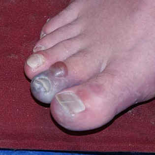
Frostbite is the damage to tissues from freezing ;The blood vessels contract and cause loss of oxygen to the affected body parts.And results formation of ice crystals within cells, rupturing the cells and leading to cell death.
It most commonly affects areas that are further away from the body core and have less blood flow. These include your feet, hands, nose, and ears.

There are three degrees of cold injury: frostnip, superficial frostbite, and deep frostbite.

The affected skin may be slightly flushed. The skin changes to white or grayish yellow as the frostbite develops. Pain is sometimes felt early but subsides later. Often there is NO pain; the part being frostbitten simply feels intensely cold and numb.

In superficial frostbite, there will be an area that looks white or grayish and the surface skin will feel hard but the underlying tissue will be soft. With deeper involvement, large blisters appear on the surface, as well as in underlying tissue, and the affected area is hard, cold and insensitive. Destruction of the entire thickness of the skin will necessitate skin grafting and will constitute a medical emergency, because gangrene may result from loss of blood supply to the injured part.

CLUBBLING the procces

It is described clinically as a bulbous uniform swelling of the soft tissue of the terminal phalanx of a digit with subsequent loss of the normal angle between the nail and the nail bed.

Since Hippocrates first described digital clubbing in patients with empyema, digital clubbing has been associated with various underlying pulmonary, cardiovascular, neoplastic, infectious, hepatobiliary, mediastinal, endocrine, and gastrointestinal diseases. Finger clubbing also may occur, without evident underlying disease


CLUBBING CAUSES:..................CLUBBING
    A 62-year-old female is brought to the emergency room by her husband with complaints of shortness of breath. Which of the following physical findings would be the most reliable indicator that she is experiencing heart failure?
    a) A third heart sound (S3)
    b) A fourth heart sound (S4)
    c) Ascites
    d) Orthopnea

    Wednesday, March 30, 2011

    Terms of Position, Direction and the main planes of human body

    The Diagram below shows the chief terms of position and direction and the main planes of reference in the body.

    *A convention whereby the body is erect, with the head, eyes, and toes directed forward and the upper limbs by the side and held so that the palms of the hands face forward "unlike the figure at left " . It is often necessary, however, to describe the position of the viscera also in the recumbent posture, because this is a posture in which patients are frequently examined clinically. 

    *The median plane is an imaginary vertical plane of section that passes longitudinally through the body and divides it into right and left halves. The median plane intersects the surface of the front and back of the body at what are called the anterior and posterior median lines. It is a common error, however, to refer to the" midline" when the median plane is meant.

    Any vertical plane through the body that is parallel with the median plane is called a sagittal plane. The sagittal planes are named after the sagittal suture of the skull, to which they are parallel. The term "parasagittal" is redundant: anything parallel with a sagittal plane is still sagittal.

    The term horizontal plane refers to a plane at a right angle to both the median and coronal planes: it separates the body into superior and inferior parts. This is often termed an axial plane, particularly in radiology.

    The term transverse means at a right angle to the longitudinal axis of a structure. Thus, a transverse section through an artery is not necessarily horizontal. A transverse section through the hand is horizontal, whereas a transverse section through the foot is coronal !!

    The suffix "-ad" is sometimes added to a positional term to indicate the idea of motion. Thus, cephalad means proceeding toward the head. Such terms are useful occasionally in describing growth processes, but their application is best limited.


    the Stomach:Arterial supply

    The arteries that supply the stomach are branches of the celiac trunk . This is the first unpaired branch of the abdominal aorta, arising just after the aorta passes behind the diaphragm.
    The branches of the celiac artery are three:

    1. left gastric
    2. splenic
    3. common hepatic


    Good Mnemonics for Thenar and hypothenar muscles

    FAO (Flex, Oppose, Abduct)

    thenar:
    Flexor pollicis brevis, Opponens pollicis, Abductor pollicis brevis. 


    hypothenar:
    Flexor digiti minimi, Abductor digiti minimi, Opponens digiti minimi.

    Diagram of Anterior anatomical relations of both kidney

    The kidneys are retroperitoneal organs that are located in the perirenal retroperitoneal space with a longitudinal diameter of 10–12 cm and a latero-lateral diameter of 3–5 cm and a weight of 250–270 g.
    In the supine position, the medial border of the normal kidney is much more anterior than the lateral border, The upper pole of each kidney is situated more posteriorly than the lower pole.

    The right kidney,  anteriorly :
    has a relation with the inferior surface of the liver with peritoneal interposition,and with the second portion of the duodenum without any peritoneal interposition since the second portion of the duodenum is retroperitoneal .
    The left kidney, anteriorly :
    has a relation with the pancreatic tail, the spleen, the stomach, the ligament of Treitz and small bowel, and with the left colic lexure and left colon . Over the left kidney, there are two important peritoneal relections, one vertical corresponding to the spleno-renal ligament (connected to
    the gastro-diaphragmatic and gastrosplenic ligaments) and one horizontal corresponding to the transverse mesocolon.


    Complete neuron cell diagram


    Neurons can respond to stimuli and conduct impulses because a membrane potential is established across the cell membrane. In other words, there is an unequal distribution of ions (charged atoms) on the two sides of a nerve cell membrane. This can be illustrated with a voltmeter:

    With one electrode placed inside a neuron and the other outside, the voltmeter is 'measuring' the difference in the distribution of ions on the inside versus the outside. And, in this example, the voltmeter reads -70 mV (mV = millivolts).
    In other words, the inside of the neuron is slightly negative relative to the outside. This difference is referred to as the Resting Membrane Potential

    ACC Atlas of Pathophysiology


    Featuring 450 large full-color illustrations, this comprehensive atlas shows how more than 200 disorders can disrupt the human body’s equilibrium. It is designed to help healthcare professionals visualize disease processes, understand the rationales for clinical interventions, and explain to patients how diseases develop and progress.

    Introductory chapters illustrate basic pathophysiologic concepts including cells, cancer, infection, genetics, and fluids and electrolytes. Twelve chapters organized by body system cover all major diseases, with illustrations, charts, and brief text on causes, pathophysiology, signs, symptoms, diagnostic tests, and treatment.

    This Third Edition includes eight new disease entries and updated information throughout. A new icon, Complications, highlights the typical progression of untreated disease.

    SLUG BAM: Muscarinic effects of Acetylcholine

    Acetylcholine acts on two vastly different classes of receptors - nicotinic receptors (with two subtypes, one at the neuromuscular junction of skeletal muscle, the other within ganglia and the CNS), and muscarinic receptors (widely distributed within both peripheral and central nervous systems).

    **Muscarinic effects SLUG BAM:
    • Salivation/ Secretions/ Sweating
    • Lacrimation
    • Urination
    • Gastrointestinal upset
    • Bradycardia/ Bronchoconstriction/ Bowel movement
    • Abdominal cramps/ Anorexia
    • Miosis

    Neural transmission in the nodes of Ranvier

    The nodes of Ranvier increase the efficiency of neural transmission by means of which of the following?

    • a.Decelerating the closing of Na -gated channels
    • b.Enhancing myelination of the internodal segment
    • c.Sequestration of Na entry into the axon
    • d.Multiple firings due to local ionic currents around the node
    • e.Decreasing threshold for the action potential

     The answer is  (c ).
    The nodes of Ranvier increase the efficiency of nodal conduction because of restriction (sequestration) of energy–dependent Na influx to the node. The nodes of Ranvier represent
    the space between adjacent units of myelination.
    This area is bare in the CNS, whereas in the PNS the axons in the nodes are partially covered by the cytoplasmic tongues of adjacent Schwann cells. Most of the Na -gated channels are located in the bare areas. Therefore, spread of depolarization from the nodal region along the axon occurs until it reaches the next node.
    This is often described as a series of jumps from node to node, or saltatory conduction.


    Tuesday, March 29, 2011

    The Physiology of Stress in human

    the stress respond

    In stress (as in threatening situations that we are unable to cope with) messages are carried along neurones from the cerebral cortex (where the thought processes occur) and the limbic system to the Hypothalamus.

    The Anterior Hypothalamus produces sympathetic arousal of the Autonomic Nervous System (ANS). The ANS is an automatic system that controls the heart, lungs, stomach, blood vessels and glands. Due to its action we do not need to make any conscious effort to regulate our breathing or heart beat. The ANS consists of : the sympathetic nervous system and the parasympathetic nervous system. Essentially, the parasympathetic nervous system conserves energy levels. It increases bodily secretions such as tears, gastric acids, mucus and saliva which help to defend the body and help digestion. Chemically, the parasympathetic system sends its messages by a neurotransmitter acetylcholine which is stored at nerve endings.

    Unlike the parasympathetic nervous system which aids relaxation, the sympathetic nervous system prepares the body for action. In a stressful situation, it quickly does the following:

    * Increases strength of skeletal muscles
    * Decreases blood clotting time
    * Increases heart rate
    * Increases sugar and fat levels
    * Reduces intestinal movement
    * Inhibits tears, digestive secretions.
    * Relaxes the bladder
    * Dilates pupils
    * Increases perspiration
    * Increases mental activity
    * Inhibits erection/vaginal lubrication
    * Constricts most blood vessels but dilates those in heart/leg/arm muscles

    The main sympathetic neurotransmitter is noradrenaline which is released a the nerve endings. The stress response also includes the activity of the adrenal, pituitary and thyroid glands.

    The two adrenal glands are located one on top of each kidney. the adrenal medulla is connected to the sympathetic nervous system by nerves. Once the latter system is in action it instructs the adrenal medulla to produce adrenaline and noradrenaline (catecholamines) which are released into the blood supply. The adrenaline prepares the body for flight and the noradrenaline prepares the body for fight. They increase both the heart rate, and the pressure at which the blood leaves the heart; they dilate bronchial passages and dilate coronary arteries; skin blood vessels constrict and there is an increase in metabolic rate. Also gastrointestinal system activity reduces which leads to a sensation of butterflies in the stomach.

    Lying close to the hypothalamus in the brain the pituitary gland. In a stressful situation, the anterior hypothalamus activates the pituitary. The pituitary releases adrenocorticotrophic hormone (ACTH) into the blood which then activates the outer part of the adrenal gland, the adrenal cortex. This then synthesises cortisol which increases arterial blood pressure, mobilises fats and glucose from the adipose (fat) tissues, reduces allergic reactions, reduces inflammation and can decrease lymphocytes that are involved in dealing with invading particles or bacteria. Consequently, increased cortisol levels over a prolonged period of time lowers the efficiency of the immune system. The adrenal cortex releases aldosterone which increases blood volume and subsequently blood pressure. Unfortunately, prolonged arousal over a period of time due to stress can lead to essential hypertension.
    the pituatary
    also releases thyroid stimulating hormone which stimulates the thyroid gland, to secrete thyroxin. Thyroxin increases the metabolic rate, raises blood sugar levels, increases respiration/heart rate/blood pressure/and intestinal motility. Increased intestinal motility can lead to diarrhoea. (It is worth noting that an over-active thyroid gland under normal circumstances can be a major contributory factor in anxiety attacks. This would normally require medication.)

    The pituitary also releases oxytocin and vasopressin which contract smooth muscles such as the blood vessels. Oxytocin causes contraction of the uterus. Vasopressin increases the permeability of the vessels to water therefore increasing blood pressure. It can lead to contraction of the intestinal musculature.

    for many people they perceive everyday of their life as stressful. Unfortunately, the prolonged effect of the stress response is that the body's immune system is lowered and blood pressure is raised which may lead to essential hypertension and headaches. The adrenal gland may malfunction which can result in tiredness with the muscles feeling weak; digestive difficulties with a craving for sweet, starchy food; dizziness; and disturbances of sleep.

    Highest oxygen content in Fetal blood

    Fetal blood from the placenta is about 80% oxygenated. However, mixture with unoxygenated blood at various points reduces the oxygen content.Which of the following fetal vessels contains blood with the highest oxygen content?
    A.Abdominal aorta
    B. Common carotid arteries
    C.Ductus arteriosus
    D.Pulmonary artery
    E.Pulmonary vein.
    the ANSWER B
    Blood from the placenta in the umbilical cord is about 80% oxygenated. Mixture with unoxygenated blood from the vitelline veins and the inferior vena cava reduces the oxygen content somewhat. However, this stream with relatively high oxygen content is directed by the valve of the inferior vena cava directly through the foramen ovale into the left atrium. This prevents admixture with oxygen-depleted blood entering the right atrium from the superior vena cava.

    Thus, the oxygen-saturated blood entering the left ventricle and pumped into the aortic arch, subclavian arteries, and common carotid arteries has the highest oxygen content. The oxygen depleted blood from the superior vena cava is directed into the right ventricle and then to the pulmonary trunk. Although a small portion of this flow passes through the lungs (where any residual oxygen is extracted by the tissue of the non-respiring lung), most is shunted into the thoracic aorta via the ductus arteriosus and thereby lowers the oxygen content of that vessel. This occurs distal to the origins of the carotid arteries and ensures that the rapidly developing brain has the best oxygen supply. The pattern of blood supply in the fetus and the changes that occur at birth are shown in the following figures.

    Synovial joint:Blood and nerve supply

    The blood supply of a synovial joint is derived from the arteries sharing in the anastomosis around the joint.And the nerve supply of a synovial joint is derived from the muscles which act on the joint ,best expressed by Hilton in 1863:
    "The same trunks of nerves, whose branches supply the groups of muscles moving a joint, furnish also a distribution of nerves to the skin over the insertions of the same muscles; and what at this moment more especially merits our attention-the interior of the joint receives its nerves from the same source."

    proprioceptive fibers endings in the capsule and ligaments are very sensitive to position and movement. Their central connections are such that they are concerned with the reflex control of posture and locomotion and the detection of position and movement.


    This diagram shows the artery supplying the epiphysis, joint capsule, and synovial membrane. The nerve that contains1. sensory (mostly pain) fibers from the capsule and synovial membrane,2. autonomic (postganglionic sympathetic) fibers to blood vessels,3. sensory (pain) fibers from the adventitia of blood vessels.